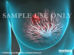
An ultrasound-guided breast biopsy uses sound waves to help locate a lump or abnormality and remove a tissue sample for examination under a microscope. It is less invasive than surgical biopsy, leaves little to no scarring and does not involve exposure to ionizing radiation.
Tell your doctor about any recent illnesses or medical conditions and whether you have any allergies, especially to anesthesia. Discuss any medications you’re taking, including herbal supplements and aspirin. You will be advised to stop taking aspirin or blood thinner three days before your procedure. Leave jewelry at home and wear loose, comfortable clothing. You may be asked to wear a gown. If you are to be sedated, plan to have someone drive you home afterward.
What is Ultrasound-Guided Breast Biopsy?
Lumps or abnormalities in the breast are often detected by physical examination, mammography, or other imaging studies. However, it is not always possible to tell from these imaging tests whether a growth is benign or cancerous.
A breast biopsy is performed to remove some cells from a suspicious area in the breast and examine them under a microscope to determine a diagnosis. This can be performed surgically or, more commonly, by a radiologist using a less invasive procedure that involves a hollow needle and image-guidance. Image-guided needle biopsy is not designed to remove the entire lesion.
Image-guided biopsy is performed by taking samples of an abnormality under some form of guidance such as ultrasound, MRI or mammographic guidance.
In ultrasound-guided breast biopsy, ultrasound imaging is used to help guide the radiologist's instruments to the site of the abnormal growth.
What are some common uses of the procedure?
An ultrasound-guided breast biopsy can be performed when a breast ultrasound shows an abnormality such as:
- a suspicious solid mass
- a distortion in the structure of the breast tissue
- an area of abnormal tissue change
There are times when your doctor may decide that ultrasound guidance for biopsy is appropriate even for a mass that can be felt.
Ultrasound guidance is used in four biopsy procedures:
- fine needle aspiration (FNA), which uses a very small needle to extract fluid or cells from the abnormal area.
- core needle (CN) which uses a large hollow needle to remove one sample of breast tissue per insertion.
- vacuum-assisted device (VAD) which uses a vacuum powered instrument to collect multiple tissue samples during one needle insertion.
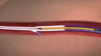
- wire localization, in which a guide wire is placed into the suspicious area to help the surgeon locate the lesion for surgical biopsy.
- How should I prepare?
You should wear comfortable, loose-fitting clothing for your ultrasound exam. You may need to remove all clothing and jewelry in the area to be examined.
You may be asked to wear a gown during the procedure.
Prior to a needle biopsy, you should report to your doctor all medications that you are taking, including herbal supplements, and if you have any allergies, especially to anesthesia. Your physician may advise you to stop taking aspirin or a blood thinner three days before your procedure.
Also, inform your doctor about recent illnesses or other medical conditions.
You may want to have a relative or friend accompany you and drive you home afterward. This is recommended if you have been sedated.
What does the equipment look like?
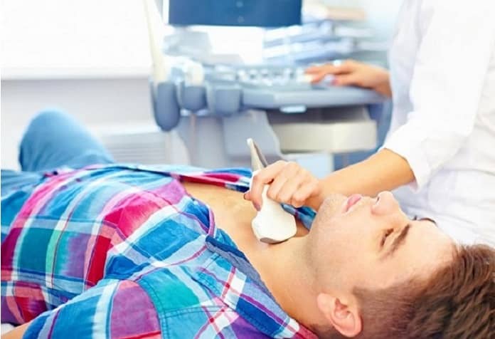
Ultrasound scanners consist of a console containing a computer and electronics, a video display screen and a transducer that is used to do the scanning. The transducer is a small hand-held device that resembles a microphone, attached to the scanner by a cord. Some exams may use different transducers (with different capabilities) during a single exam. The transducer sends out inaudible, high—frequency sound waves into the body and then listens for the returning echoes from the tissues in the body. The principles are similar to sonar used by boats and submarines.
The ultrasound image is immediately visible on a video display screen that looks like a computer or television monitor. The image is created based on the amplitude (loudness), frequency (pitch) and time it takes for the ultrasound signal to return from the area within the patient that is being examined to the transducer (the device used to examine the patient), as well as the type of body structure and composition of body tissue through which the sound travels. A small amount of gel is put on the skin to allow the sound waves to best travel from the transducer to the examined area within the body and then back again. Ultrasound is an excellent modality for some areas of the body while other areas, especially the lungs, are poorly suited for ultrasound.
One of four instruments will be used:
- A fine needle attached to a syringe, smaller than needles typically used to draw blood.
- A core needle, also called an automatic, spring-loaded needle, which consists of an inner needle connected to a trough, or shallow receptacle, covered by a sheath and attached to a spring-loaded mechanism.
- A vacuum-assisted device (VAD), a vacuum-powered instrument that uses pressure to pull tissue into the needle.
- A thin guide wire, which is used for a surgical biopsy.
Other sterile equipment involved in this procedure includes syringes, sponges, forceps, scalpels and a specimen cup or microscope slide.
How does the procedure work?
Ultrasound imaging is based on the same principles involved in the sonar used by bats, ships and fishermen. When a sound wave strikes an object, it bounces back, or echoes. By measuring these echo waves, it is possible to determine how far away the object is as well as the object's size, shape and consistency (whether the object is solid or filled with fluid).
In medicine, ultrasound is used to detect changes in appearance, size or contour of organs, tissues, and vessels or to detect abnormal masses, such as tumors.
In an ultrasound examination, a transducer both sends the sound waves and receives the echoing waves. When the transducer is pressed against the skin, it directs small pulses of inaudible, high-frequency sound waves into the body. As the sound waves bounce off internal organs, fluids and tissues, the sensitive microphone in the transducer records tiny changes in the sound's pitch and direction. These signature waves are instantly measured and displayed by a computer, which in turn creates a real-time picture on the monitor. One or more frames of the moving pictures are typically captured as still images. Short video loops of the images may also be saved.
Using an ultrasound probe to visualize the location of the breast mass, distortion or abnormal tissue change, the radiologist inserts a biopsy needle through the skin, advances it into the targeted finding and removes tissue samples. If a surgical biopsy is being performed, ultrasound may be used to guide a wire directly into the targeted finding to help the surgeon locate the area for excision. With continuous ultrasound imaging, the physician is able to view the biopsy needle or wire as it advances to the location of the lesion in real-time.
How is the procedure performed?
Image-guided, minimally invasive procedures such as ultrasound-guided breast biopsy are most often performed by a specially trained radiologist.
Breast biopsies are usually done on an outpatient basis.
You will be positioned lying face up on the examination table or turned slightly to the side.
A local anesthetic will be injected into the breast to numb it.
Pressing the transducer to the breast, the sonographer or radiologist will locate the lesion.
A very small nick is made in the skin at the site where the biopsy needle is to be inserted.
The radiologist, monitoring the lesion site with the ultrasound probe, will insert the needle and advance it directly into the mass.
Tissue samples are then removed using one of three methods:
- In a fine needle aspiration, a fine gauge needle and a syringe withdraw fluid or clusters of cells.
- In a core needle biopsy, the automated mechanism is activated, moving the needle forward and filling the needle trough, or shallow receptacle, with 'cores' of breast tissue. The outer sheath instantly moves forward to cut the tissue and keep it in the trough. This process is repeated three to six times.
- With a vacuum-assisted device (VAD), vacuum pressure is used to pull tissue from the breast through the needle into the sampling chamber. Without withdrawing and reinserting the needle, it rotates positions and collects additional samples. Typically, eight to 10 samples of tissue are collected from around the lesion.
After this sampling, the needle will be removed.
If a surgical biopsy is being performed, a wire is inserted into the suspicious area as a guide for the surgeon.
A small marker may be placed at the biopsy site so that it can be located in the future if necessary.
Once the biopsy is complete, pressure will be applied to stop any bleeding and the opening in the skin is covered with a dressing. No sutures are needed.
A mammogram may be performed to confirm that the marker is in the proper position.
This procedure is usually completed within an hour.
What will I experience during and after the procedure?
You will be awake during your biopsy and should have little discomfort. Most women report little or no pain and no scarring on the breast.
When you receive the local anesthetic to numb the skin, you will feel a slight pin prick from the needle. You may feel some pressure when the biopsy needle is inserted.
The area will become numb within a short time.
You must remain still while the biopsy is performed.
As tissue samples are taken, you may hear clicks or buzzing sounds from the sampling instrument.
If you experience swelling and bruising following your biopsy, you may be instructed to take an over-the-counter pain reliever and to use a cold pack. Temporary bruising is normal.
You should contact your physician if you experience excessive swelling, bleeding, drainage, redness or heat in the breast.
If a marker is left inside the breast to mark the location of the biopsied lesion, it will cause no pain, disfigurement or harm.
You should avoid strenuous activity for 24 hours after the biopsy. After that period of time, you will usually be able to resume normal activities.
Who interprets the results and how do I get them?
A pathologist examines the removed specimen and makes a final diagnosis. Depending on the facility, the radiologist or yourreferring physician will share the results with you. The radiologist will also evaluate the results of the biopsy to make sure that the pathology and image findings are appropriate.
Follow-up examinations may be necessary, and your doctor will explain the exact reason why another exam is requested. Sometimes a follow-up exam is done because a suspicious or questionable finding needs clarification with additional views or a special imaging technique. A follow-up examination may also be necessary so that any change in a known abnormality can be monitored over time. Follow-up examinations are sometimes the best way to see if treatment is working or if an abnormality is stable or changes over time.
What are the benefits vs. risks?
Benefits
- The procedure is less invasive than surgical biopsy, leaves little or no scarring and can be performed in less than an hour.
- Ultrasound imaging uses no ionizing radiation.
- Ultrasound-guided breast biopsy reliably provides tissue samples that can show whether a breast lump is benign or malignant.
- Compared with stereotactic breast biopsy, the ultrasound method is faster and avoids the need for ionizing radiationexposure.
- With ultrasound it is possible to follow the motion of the biopsy needle as it moves through the breast tissue.
- Ultrasound-guided breast biopsy is able to evaluate lumps under the arm or near the chest wall, which are hard to reach with stereotactic biopsy.
- Ultrasound-guided biopsy is less expensive than other biopsy methods, such as open surgical biopsy or stereotactic biopsy.
- Recovery time is brief and patients can soon resume their usual activities.
Risks
- There is a risk of bleeding and forming a hematoma, or a collection of blood at the biopsy site. The risk, however, appears to be less than one percent of patients.
- An occasional patient has significant discomfort, which can be readily controlled by non-prescription pain medication.
- Any procedure where the skin is penetrated carries a risk of infection. The chance of infection requiring antibiotic treatment appears to be less than one in 1,000.
- Depending on the type of biopsy being performed or the design of the biopsy machine, a biopsy of tissue located deep within the breast carries a slight risk that the needle will pass through the chest wall, allowing air around the lung that could cause the lung to collapse. This is an extremely rare occurrence.
- What are the limitations of Ultrasound-Guided Breast Biopsy?
Breast biopsy procedures will occasionally miss a lesion or underestimate the extent of disease present. If the diagnosis remains uncertain after a technically successful procedure, surgical biopsy will usually be necessary.
The ultrasound-guided biopsy method cannot be used unless the lesion can be seen on an ultrasound exam. Clustered calcifications are not shown as clearly with ultrasound as with x-rays.
Very small lesions may be difficult to target accurately by ultrasound-guided core biopsy.


Leave a Reply
Want to join the discussion?Feel free to contribute!